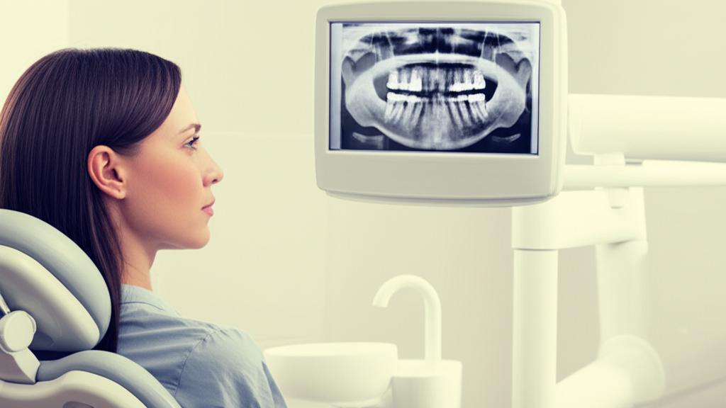
I remember a patient, let’s call her Sarah, who came in for her regular check-up. On the surface, everything looked pretty good. No major complaints, just a little “awareness” she said, around one of her back teeth. Nothing a visual check could pinpoint. But then we took a couple of routine dental X-rays. And there it was – a sneaky little cavity starting to form between two teeth, right where we couldn’t see. Without that X-ray, it would have grown, no doubt, and caused her a lot more trouble down the line. That’s the power of these images.
So, what exactly are dental X-rays? Think of them as special photographs, radiographs as we call them in the biz, that let us see inside your teeth and jaws. There’s a lot going on under the surface that we just can’t see with our eyes alone – your jawbone, nerves, sinuses, and the roots of your teeth. These X-rays help us get the full picture of your oral health.
How Do Dental X-rays Work Their Magic?
It’s pretty clever, really. Like X-rays for other parts of your body, dental X-rays use a very focused beam of electromagnetic radiation. This beam can pass through your softer tissues, like your cheeks and gums, but it’s absorbed more by denser things like your teeth and bones. This creates an image, showing us those harder structures.
You might hear about traditional X-rays, which use film, or digital ones. Most clinics, mine included, have moved to digital dental X-rays. The big plus? They use significantly less radiation – we’re talking 80% to 90% less than the older machines. That’s a big win for safety.
What Can We Spot with Dental X-rays?
These images are like detective tools for us. With dental X-rays, we can often see:
- Cavities, especially those little areas of decay tucked away between your teeth.
- Decay hiding beneath existing fillings. Sometimes old fillings can have issues underneath.
- Bone loss in your jaw, which can be a sign of gum disease.
- Areas of infection that might be brewing.
- The position of teeth that haven’t quite made it through the gums yet (unerupted teeth) or teeth that are stuck (impacted teeth).
- Abscessed teeth – that’s an infection right at the root of your tooth or between the gum and the tooth. Ouch.
- Cysts and, less commonly, some types of tumors.
We also use X-rays to plan treatments. Thinking about dental implants, braces, or dentures? X-rays are crucial. And after procedures like dental bone grafts or root canal therapy, they help us check that everything is healing up just right.
Getting Your Dental X-rays: What to Expect
The X-ray Process: Quick and Easy
If you’re due for X-rays, it’s a pretty straightforward process. First, we’ll drape a lead apron over your chest and lap. Sometimes, we’ll also use a thyroid collar that fits around your neck. These are just to protect you from any unnecessary radiation. Safety first, always.
Then, depending on the type of X-ray, you’ll either sit in the dental chair or stand by the X-ray machine. A dental assistant or hygienist will carefully place a small film or a digital sensor in your mouth. You might be asked to bite down gently. Then, they’ll step out for a second, press a button, and… click! It’s over in an instant. The main thing is to try and hold still for that brief moment.
Exploring the Different Types of Dental X-rays
Not all X-rays are the same; we use different types for different reasons. They mainly fall into two groups:
- Intraoral X-rays: This means the film or sensor is inside your mouth.
- Extraoral X-rays: For these, the film or sensor is outside your mouth.
Intraoral X-rays: A Closer Look Inside
These are the ones you’re probably most familiar with:
- Bitewing X-rays: You’ll bite down on a little tab for these. They show the crowns of your upper and lower teeth in one specific area of your mouth. Perfect for finding decay between teeth or changes just below the gum line. They don’t usually show the full roots, though.
- Periapical X-rays: This type gives us a view of the entire tooth, from the chewing surface (the crown) all the way down to the tip of the root and the bone around it. We use these to check for decay, gum disease, bone loss, or any other issues with a specific tooth.
- Occlusal X-rays: These are a bit larger and show almost the full arch of your teeth in either the upper or lower jaw. They help us see the floor or roof of your mouth. They’re useful for finding fractured teeth, impacted teeth, or checking the development of children’s teeth. They can also help spot cysts, abscesses, or even jaw fractures.
Extraoral X-rays: The Bigger Picture
Sometimes we need to see more than just a few teeth:
- Panoramic X-rays: This is the one where the machine moves around your head. It gives us a single, wide view of your entire mouth – all your teeth, upper and lower jaws, jaw joints (TMJ), nerves, and sinuses. It’s a fantastic overview.
- Cephalometric X-rays: You’ll see this one taken from the side of your head. It shows us how your teeth relate to your jaw and profile. Orthodontists (the dentists who specialize in straightening teeth and correcting bites) use these a lot when planning for braces.
- Cone beam CT scan (CBCT): This is a more advanced type, giving us 3D images. A dental CT scan provides incredibly detailed views of your teeth, soft tissues, nerve pathways, and bone, all in a single scan. We might use this for complex cases, like planning for dental implant placement, or if we suspect tumors or facial fractures.
Let’s Talk Safety: Are Dental X-rays Okay?
This is a question I hear a lot, and it’s a good one. The amount of radiation you receive from a dental X-ray is actually very small. To put it in perspective, a full set of dental X-rays exposes you to about the same amount of radiation you’d naturally absorb from the environment (like from the sun or even certain building materials) over a few days. Digital X-rays, which we use, have even lower levels.
Now, in extremely large doses, radiation can be harmful, potentially increasing cancer risk. That’s why we’re always careful and only take X-rays when they’re truly necessary to diagnose or treat a problem. We always weigh the benefits of getting a clear picture of your oral health against the tiny risk.
After the X-ray: What’s Next?
How Often Do You Need Dental X-rays?
There’s no one-size-fits-all answer, but here’s a general guide. If your teeth and gums are generally healthy, we typically recommend getting bitewing X-rays about once every 6 to 18 months. A full set, including periapical or a panoramic X-ray, might be less frequent, maybe every few years.
However, if you have ongoing issues like recurrent tooth decay, active gum disease, or other specific concerns, we might need to take them more often to keep a close eye on things. It really depends on your individual situation.
Your Questions Answered
Can I Say No to Dental X-rays?
Absolutely. As a patient, you always have the right to refuse any medical procedure, including dental X-rays. However, it’s important to understand why your dentist is recommending them. Without X-rays, we might miss problems that aren’t visible on the surface. It’s like trying to understand what’s wrong with a car engine without looking under the hood. Most dentists feel they can’t provide the best care, or sometimes any care, without them because they can’t fully assess your condition. If you have concerns about radiation, please talk to us. We can explain the specific reasons for the X-ray and discuss any alternatives, if available.
Can a Dental X-ray Spot Cancer?
Dental X-rays can sometimes show signs of certain types of oral cancer, particularly if cancer has started in or spread to your jawbone. However, they can’t detect all types of mouth cancer, especially in the early stages or in soft tissues. That’s why regular oral cancer screenings, which are a visual and manual check of your mouth, are so crucial during your dental visits.
What About Dental X-rays During Pregnancy?
This is another common concern. Generally, dental X-rays are considered safe during pregnancy, especially with the lead apron and thyroid collar. Both the American Dental Association and the American Pregnancy Association have confirmed the risk to the developing baby is very low. That said, unless it’s an emergency (like a severe infection or pain that needs immediate diagnosis), many dentists, myself included, prefer to postpone routine X-rays until after the baby is born. If an X-ray is absolutely necessary, we take every precaution. If you’re pregnant or think you might be, always let your dental team know.
Key Things to Remember About Dental X-rays
Here’s a quick rundown of what’s most important:
- Dental X-rays are essential tools that let us see hidden problems in your teeth and jaws.
- They help us find cavities, infections, bone loss, and plan treatments like implants or braces.
- Digital X-rays use very low levels of radiation, comparable to everyday environmental exposure.
- We always use protective gear like lead aprons.
- How often you need them depends on your individual oral health.
- Don’t hesitate to ask questions if you have any concerns about dental X-rays.
You’re in good hands. We always want to make sure we have the clearest possible picture of your health so we can take the best care of you and your smile.


