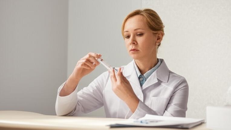
It’s that moment, isn’t it? You feel a new lump, or maybe I’ve pointed out an area on a scan that looks a little different. Your mind might start racing. That’s perfectly normal. One of the first steps we often take to get clear answers is a procedure called fine-needle aspiration, or FNA for short. It sounds a bit technical, but it’s actually a pretty straightforward way for us to understand what’s going on beneath the surface.
So, What Exactly is Fine-Needle Aspiration (FNA)?
Think of fine-needle aspiration as a very precise way to gather a tiny sample. We use a very thin – “fine” – needle attached to a syringe to gently draw out a small collection of cells, a bit of tissue, or some fluid from that suspicious spot. It’s one of the least invasive ways to get a sample, which is why we often consider it.
Once we have that sample, it’s sent off to a cytology lab. There, a cytotechnologist (a specialist trained to screen cells) prepares it, and then a pathologist (a doctor who diagnoses diseases by looking at cells and tissues under a microscope) examines it closely. They’re looking for clues about what those cells are and whether they’re behaving normally.
FNA vs. Core Biopsy: What’s the Difference?
You might hear about other types of biopsies, like a core biopsy. Both are needle biopsies, but there are key differences.
- The needle for a core biopsy is a bit wider than the one for an FNA. This means it collects a larger piece of tissue, which can sometimes give us more detailed information.
- An FNA is usually just the needle. A core biopsy might occasionally need a tiny nick in the skin to insert the needle.
We decide which type of biopsy is best based on where the lump is, its size, and what kind of information we need. For many situations, an FNA is an excellent and less disruptive starting point.
Why Would I Need an FNA?
We use fine-needle aspiration for a couple of main reasons:
Getting a Diagnosis
This is the most common reason. An FNA can help us figure out what’s causing a lump or an unusual area. By looking at the cells, the pathologist can often tell us if it’s:
- Cancerous: Identifying cancer cells.
- An infection: Sometimes bacteria can cause lumps, like in bursitis or septic arthritis.
- Noncancerous (benign): Things like cysts (fluid-filled sacs), fibroadenomas (common benign breast lumps), or lipomas (fatty lumps).
We can do an FNA on many parts of the body. It’s very commonly used for new lumps in the:
- Breast (like draining a breast cyst)
- Thyroid (to check thyroid nodules)
- Lymph nodes (those little glands that can swell up)
- Skin
Sometimes, the lump is deeper inside your body. In those cases, we might use an imaging guide, like an ultrasound or a CT scan, to help us see exactly where to place the needle. This allows us to get samples from places like your:
- Pancreas
- Lungs
- Liver
- Other organs in your chest or abdomen
FNAs can also be useful if cancer has spread (metastatic cancer). The cells can sometimes tell us about genetic markers that help guide which treatments might work best.
For Treatment
Less commonly, an FNA can also be a form of treatment. For example, if you have a painful cyst or an abscess (a collection of pus), we can use the needle to drain the fluid. This can relieve discomfort and help the area heal. We might also use it for seromas, which are fluid collections that can happen after surgery.
What to Expect: The FNA Procedure
Knowing what’s going to happen can make any procedure feel less daunting.
How Do I Get Ready?
Usually, there’s not much special preparation needed for an FNA. Just wear comfortable clothing. If there are any specific instructions, like about medications you’re taking (especially blood thinners), we’ll go over those with you well beforehand.
During the FNA
The FNA is typically done right here in the clinic or as an outpatient procedure at the hospital. Here’s a general idea of what happens:
- You’ll either sit or lie down, depending on where the sample is being taken from.
- We’ll clean the skin over the area really well.
- Often, we’ll numb the spot with a little local anesthetic. You might feel a quick pinch from that, but it helps make the FNA itself more comfortable. You could still feel a bit of pressure when the biopsy needle goes in.
- If the lump is deep, we’ll use an imaging machine (like an ultrasound) to guide the needle to the exact spot.
- Then, the fine needle is gently inserted into the lump or area. We’ll draw back on the syringe to collect the cells or fluid. This might be repeated a couple of times in slightly different spots within the lump to make sure we get a good sample. We’ll use a new needle and syringe for each pass if needed.
- Once we have what we need, the needle comes out, and we’ll put a small bandage on the spot.
The whole thing is usually quite quick!
After the FNA
The sample goes straight to the lab for the pathologist to examine. It can take a few days to get the results back. Don’t worry, we’ll call you as soon as we have them and discuss what they mean.
Are There Any Risks with FNA?
Fine-needle aspiration is generally very safe. The most common things people experience are:
- A bit of soreness at the site, like a bruise.
- Some minor bruising.
These usually fade within a day or two. An ice pack can help if you’re uncomfortable.
More serious complications are rare, but it’s good to be aware of them:
- Bleeding: There’s a small chance of bleeding, especially if you have a bleeding disorder or take blood-thinning medication. We’ll always ask about this beforehand.
- Infection: Any time the skin is broken, there’s a tiny risk of infection, but we take great care to keep everything sterile.
- Damage to nearby structures: This is very uncommon, especially when we use imaging guidance for deeper samples.
- Fistula: An abnormal connection between tissues – extremely rare.
Understanding Your FNA Results
When the results come in, we’ll sit down and talk them through. The pathologist’s report might show:
- Normal cells: No signs of anything worrying.
- Abnormal cells: This could mean a few things. Sometimes the cells look unusual but aren’t cancerous (atypical cells). We might need more tests.
- Precancerous cells: Cells that aren’t cancer yet but could become cancer over time.
- Cancerous cells: If cancer is found, the report will give us more details about the type.
- Infection: Signs of bacteria or other infectious agents.
- Insufficient sample: Sometimes, the sample might not have enough cells for a clear diagnosis. If this happens, we might need to repeat the FNA or consider a different type of biopsy. It’s frustrating, I know, but it happens.
No matter the result, we’ll discuss what it means for you and what the next steps should be.
When Should You Call Me?
After your FNA, please give us a call if you notice:
- Signs of infection: like increasing redness, warmth, swelling, pus at the needle site, or if you develop a fever.
- Significant bleeding that doesn’t stop with a little pressure.
- If the lump changes in an unexpected way.
- Any other concerns you might have.
Take-Home Message About Fine-Needle Aspiration
Here are the key things to remember about fine-needle aspiration (FNA):
- It’s a quick, minimally invasive test to get cell samples from lumps or unusual areas.
- It helps us diagnose conditions like cancer, infections, or benign growths.
- Preparation is usually simple, and the procedure itself is generally well-tolerated.
- Results help guide our next steps in your care.
- While very safe, watch for any signs of infection or unusual bleeding afterward.
Remember, if we’re suggesting an FNA, it’s because we want to get the clearest possible picture of what’s going on so we can provide you with the best care. We’ll walk through it together.
You’re not alone in this. We’re here to answer all your questions.


