The word itself – “gonioscopy” – can sound a bit intimidating, can’t it? I’ve had patients chuckle, saying it sounds like something out of a science fiction movie. But honestly, it’s a really straightforward and incredibly helpful peek inside your eye. If you’ve ever wondered how we check for certain eye conditions, especially things like glaucoma, then gonioscopy is a key part of that story. It’s a test an eye care specialist, often an ophthalmologist, uses to get a good look at the front part of your eye, specifically the drainage system. Think of it as making sure the plumbing in your eye is working just right.
What Exactly is Gonioscopy? (And Why We Do It)
So, what’s the big deal about this drainage system? Well, inside your eye, there’s a fluid-filled space called the anterior chamber. It’s right behind the clear front part (the cornea) and in front of the colored part (the iris). This chamber is full of a fluid called aqueous humor. For your eye to stay healthy, this fluid needs to drain out properly. The spot where it drains is called the drainage angle, a little ring where your iris and the white part of your eye (the sclera) meet.
If this angle is too narrow or blocked, the fluid can’t drain. Pressure builds up inside your eye – we call this intraocular pressure. And that, my friend, is how glaucoma can start. High pressure, if left unchecked, can damage the optic nerve at the back of your eye, leading to vision loss. It’s serious stuff.
Gonioscopy lets your specialist see this angle directly. It’s like using a tiny, cleverly designed periscope to look around a corner. We perform it to check for:
- Angle-closure glaucoma: Where the angle is suddenly blocked.
- Open-angle glaucoma: Where the angle is open, but drainage is still slow.
- Pseudoexfoliation syndrome: A condition that can clog the drainage system.
- Sometimes, even tiny cancers or tumors affecting the iris.
Oh, and if you’re wondering how to say it, you’ve got two options: “Go-nee-OSS-co-pee” or “Gaw-nee-OSS-co-pee.” Both work!
How does it work? The Magic of Lenses
There are two main ways we do this, using special lenses:
- Indirect gonioscopy lenses: These use mirrors. The light bounces off the mirrors, allowing the specialist to see the drainage angle. This is the most common type you’d experience in an office setting.
- Direct gonioscopy lenses: These don’t have mirrors. They magnify and bend the light a bit, so the specialist can look straight at the angle. This type is more often used in a hospital setting, sometimes during surgery.
Getting Ready for Your Gonioscopy
Good news! For most gonioscopy exams, especially the indirect kind in the clinic, you don’t usually need to do any special prep. Easy peasy.
One little thing: if you wear contact lenses, it’s always a good idea to ask your eye specialist if you should take them out for a certain period before the test. If your gonioscopy is part of a surgical procedure, then, of course, your specialist will give you very specific instructions on how to prepare.
What Happens During the Gonioscopy Test?
Alright, let’s walk through what it’s like.
If it’s Direct Gonioscopy…
This usually happens in a hospital, and often you’ll be under general anesthesia. That means you’ll be comfortably asleep while the specialist does their work.
If it’s Indirect Gonioscopy (More Common in Clinic)…
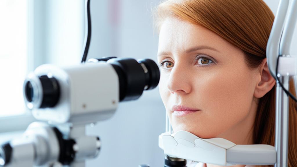
This will feel a lot like a regular slit lamp exam – you know, where you sit in a chair with your chin on a rest, looking into a microscope-like device.
- First things first, we’ll put some numbing drops in your eye. Your cornea is super sensitive, so these drops make sure you’re comfortable and don’t feel the urge to blink.
- Next, your specialist will put some wetting drops or a similar gel on the surface of the special gonioscopy lens that will touch your eye. This helps create a clear view by pushing out tiny air bubbles and allows the lens to move smoothly.
- Then, the specialist will gently place the lens on your eye, holding it with one hand. With the other hand, they’ll operate the slit lamp, which has a bright light and a magnifier.
- They’ll ask you to look straight ahead, or sometimes in specific directions. They might also do something called dynamic gonioscopy, where they use the lens to apply very gentle pressure to your eye. This helps them see if your iris is stuck to your cornea or lens (a condition called synechiae). It sounds a bit odd, but it doesn’t hurt because of the numbing.
- Once they’ve had a good look all around the edge of your iris, they’ll remove the lens. Your eye might water a bit, and that’s totally normal. We’ll have tissues ready.
Sometimes, your eyes might be dilated for this test, or the specialist might want to look before and after using dilating drops. They’ll let you know what’s needed for you.
Does it Hurt? Any Risks?
The gonioscopy test itself is very safe and shouldn’t hurt a bit, thanks to those numbing drops. You might feel a slight sensation of pressure, kind of like if you gently rub your eye, but that’s about it. The medications used (numbing or dilating drops) can sometimes have minor, temporary side effects like blurry vision or light sensitivity if dilating drops are used. Your specialist will go over anything like that with you.
After the Gonioscopy: Understanding Your Results
If you have an indirect gonioscopy in the office, your eye specialist can often tell you what they see right away, or shortly after the exam. They’ll explain if the drainage angle looks open and healthy, or if it seems a bit narrow or has any other issues.
If it was a direct gonioscopy during a procedure where you were asleep, your specialist will talk to you once you’re awake and explain their findings.
What if the Results Aren’t Normal?
If the drainage angle is narrowed, or if there’s evidence the aqueous humor isn’t draining well, don’t worry – the next step is to talk about options. Treating glaucoma early is so important because that pressure buildup can cause permanent damage to your optic nerve and affect your vision. We want to prevent that.
Sometimes, rarely, the drainage angle can be completely closed. This is called angle-closure glaucoma, and it’s a medical emergency because it can cause sudden, and often permanent, vision loss if not treated quickly. If this is suspected, your specialist will guide you on getting emergency treatment.
When to Chat with Your Eye Specialist (Or Head to the ER)
Glaucoma often develops slowly, without many early symptoms. That’s why regular eye exams are your best friend, especially every one to two years, or annually if you’re over 40. You might need more frequent checks, including gonioscopy, if you:
- Are 65 or older
- Have Type 2 diabetes
- Have a family history of glaucoma
- Have high pressure inside your eye (ocular hypertension)
- Have certain inherited eye conditions
- Use corticosteroids long-term
- Have any optic nerve damage or nerves that look a bit suspicious
- Are very nearsighted or farsighted
- Have sickle cell disease
Definitely call your eye specialist if you notice symptoms like:
- Red eyes
- Headaches (especially around the eyes)
- Nausea and vomiting (can sometimes be linked to eye pressure)
- A feeling of eye pressure or pain (usually only when pressure is very high)
- Seeing rainbow-like halos around lights
- Low vision, like tunnel vision, or new blind spots
When is it an Emergency?
Go to the ER or seek immediate medical help if you experience:
- Sudden vision loss
- Sudden, severe eye pain, especially if it comes with a headache, nausea, or vomiting.
A Quick Note: Is Gonioscopy the Whole Story?
While gonioscopy is a super valuable tool, it’s just one piece of the puzzle when we’re looking into glaucoma. Diagnosing and managing glaucoma involves looking at several factors together. And how long does it take? The actual gonioscopy part is quick, usually under five minutes. Your whole appointment might be longer if other tests are needed.
Key Things to Remember About Gonioscopy
It’s a lot to take in, I know! So here are the main takeaways:
- Gonioscopy is a safe, painless eye test to check your eye’s drainage angle.
- It’s crucial for diagnosing and monitoring conditions like glaucoma.
- Numbing drops mean you won’t feel discomfort.
- It helps your specialist see if fluid can drain properly from your eye.
- Early detection of problems like a narrow angle can save your sight.
- If you have risk factors for glaucoma, regular eye exams that may include gonioscopy are vital.
We’re In This Together
If a gonioscopy is recommended for you, please don’t hesitate to ask any questions you have. We’re here to make sure you understand every step of your eye care. You’re doin’ great just by learning more about it!


