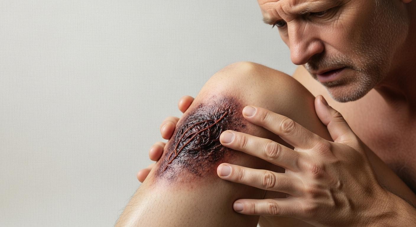
I remember a patient, Mr. Henderson, a keen gardener, who came in once with a nasty-looking cut on his leg that just wasn’t healing right. It had started to look dark, and he was worried. That worry is something I see often when the body isn’t doing what we expect. Sometimes, despite our best efforts, cells in our body tissue can die, and that’s what we call necrosis. It’s a word that sounds pretty serious, and honestly, it is. This process of necrosis can happen for a few reasons – maybe an injury, a stubborn infection, or even a disease interfering with things. Sometimes, it’s simply because blood, with all its vital oxygen and nutrients, can’t get to where it needs to go.
Think of it like a plant that doesn’t get water. The leaves wither, turn brown, and eventually, that part of the plant dies. It’s a similar idea with our tissues. Unfortunately, once tissue has undergone necrosis, we can’t bring it back to its former healthy state, though we can often remove the damaged tissue.
What Does Necrosis Look Like? The Different Patterns
When cells die from necrosis, they don’t all look the same. It’s like they leave behind different… well, signatures, you could say. Pathologists, the doctors who look at tissues under a microscope, have identified a few main patterns. It helps us understand what might be going on.
Here are the main ways these changes can appear:
- Coagulative necrosis: This is when the dead cells surprisingly keep their shape, at least for a few days. They’re firm, but they’re not alive. This often happens when blood flow or oxygen gets cut off to an area – pretty much anywhere except the brain.
- Liquefactive necrosis: With this type, the dead cells turn into a thick, goopy liquid, sometimes within hours. If there’s an infection, you might see a creamy yellow color because of pus forming. This is typical of infections or when the brain loses its oxygen supply.
- Fat necrosis: Imagine tiny cells releasing their contents, which then react with calcium in the body. This creates chalky, white spots. We see this most often with acute pancreatitis (a sudden inflammation of the pancreas), but it can also happen in breast tissue after an injury.
- Caseous necrosis: This one has a unique look – soft, white, and a bit crumbly, often described as “cheese-like.” In fact, “caseous” means just that! It’s a hallmark of tuberculosis, especially in the lungs.
- Fibrinoid necrosis: Here, the damaged area looks pink and a bit messy under the microscope. This happens when proteins from the blood (called fibrins) leak out of damaged blood vessel walls, often due to autoimmune conditions or certain infections affecting those vessels.
- Gangrenous necrosis: This is a term you might have heard. It often involves skin turning black and starting to decay. It’s usually caused by a severe lack of blood flow, commonly in the legs, but fingers and arms can be affected too. We call this gangrene.
Where Can Necrosis Happen? Understanding the Types
Necrosis isn’t just one thing; it can show up in different parts of the body, affecting bones, skin, and even our internal organs.
Here are some of the specific types we encounter:
- Avascular necrosis (Osteonecrosis): You might hear this called osteonecrosis, aseptic necrosis, or bone necrosis. It all means the same thing: bone tissue dies because its blood supply gets cut off. Bones can then weaken and break down. The hip is a common spot for this – we call it hip necrosis.
- Osteonecrosis of the jaw (ONJ): This is a specific type of avascular necrosis affecting the jawbone. It can be quite painful, and sometimes you might see pus.
- Pancreatic necrosis: This is a serious complication if someone has acute pancreatitis. If the pancreas loses its blood supply, its tissue can die. This is risky because it can get infected, and that infection can spread, leading to a body-wide problem called sepsis.
- Fat necrosis of the breast: This might sound alarming, but it’s usually a noncancerous (benign) issue. If fatty breast tissue gets damaged (say, from an injury or surgery), sometimes, instead of forming normal scar tissue, some fat cells die and create a little pocket of oily fluid. We call this an oil cyst.
- Acute tubular necrosis (ATN): This affects the kidneys. The kidneys have tiny tubes, or tubules, that filter our blood. If these tubule cells get damaged, it can lead to acute kidney failure.
- Radiation necrosis: This is a less common issue, but it can happen after high-dose radiation therapy, especially to the brain, head, or neck. Sadly, it can cause permanent death of brain tissue.
- Renal papillary necrosis: Another kidney-related problem. The renal papillae are like little funnels where urine collects before heading to the bladder. If these die, it can cause significant kidney trouble.
- Skin necrosis (Gangrene): As mentioned with gangrenous necrosis, this is when skin tissue dies due to lack of blood flow or sometimes a severe bacterial infection. Fingers, toes, hands, and feet are common spots.
- Spider bite necrosis: Thankfully rare! But bites from certain spiders, like the recluse spider, contain venom that can cause skin tissue to decay around the bite.
- Pulp necrosis: This happens inside a tooth. The soft center of your tooth, the pulp, has blood vessels and nerves. If a tooth gets badly decayed or injured, this pulp can get infected and die.
When We Talk About How Cells Die: Necrosis vs. Apoptosis
You might hear another term, apoptosis, and wonder if it’s the same thing. It’s not, really.
- Apoptosis is like the body’s natural, programmed way for cells to die when they’re old or no longer needed. It’s a tidy process, important for keeping everything in balance. Think of it as scheduled maintenance.
- Necrosis, on the other hand, is accidental cell death. It’s caused by some kind of damage – injury, infection, lack of blood flow. It’s more like an unexpected system failure.
Because necrosis is unplanned and often harmful, it usually needs our attention and treatment, while apoptosis rarely does.
A Quick Word on Fillers and TNF
Just a couple of other related things I get asked about:
Some folks worry about necrosis after dermal filler injections. These are cosmetic treatments, like hyaluronic acid injections, used to plump skin. They’re generally safe, but in rare cases, necrosis can happen if the filler accidentally blocks a blood vessel. Signs can show up right away or a few hours later and include more pain than expected, tenderness, or skin changing color (looking white, blotchy red, or even bluish). If you ever see this after a filler, it’s crucial to get it checked out immediately.
Then there’s something called Tumor Necrosis Factor (TNF). This is a protein our immune system uses. When certain white blood cells spot an infection, they release TNF to call other immune cells to action, causing inflammation to fight the problem. Interestingly, researchers are also looking into whether TNF can cause necrosis in some cancer cells, so it’s being studied for cancer treatment. Pretty clever, how our bodies work, eh?
What We Do If Necrosis Is Suspected
If we suspect necrosis, our first steps are to figure out what’s causing it and how much tissue is affected. This might involve:
- A good look at the area.
- Blood tests to check for infection or other underlying issues.
- Sometimes imaging tests like X-rays, CT scans, or MRIs can help us see what’s happening deeper inside.
- A biopsy, where a small sample of the affected tissue is taken and looked at under a microscope by a pathologist, can confirm necrosis and help identify the type.
Treatment really depends on the cause, location, and extent of the necrosis. The main goals are usually:
- Treating the underlying cause: If it’s an infection, we’ll use antibiotics. If it’s poor blood flow, we might look at ways to improve circulation.
- Debridement: This means removing the dead tissue. This is important because dead tissue can prevent healing and become a breeding ground for infection.
- Preventing further damage and infection.
- Managing pain.
- In some cases, like with gangrene, more extensive surgery, or even amputation, might be necessary to save a limb or a life.
We’ll always discuss all the options available for you or your loved one.
Key Things to Remember About Necrosis
It’s a lot to take in, I know. So, here are the main points:
- Necrosis is the death of cells and tissues in your body, often due to injury, infection, or lack of blood supply.
- It can look different depending on the cause and type (e.g., coagulative, liquefactive, gangrenous).
- Necrosis can affect various parts of the body, including bones (avascular necrosis), skin (gangrene), and internal organs.
- It’s different from apoptosis, which is normal, programmed cell death.
- Early recognition and treatment are key to managing necrosis and preventing serious complications.
- Treatment focuses on removing dead tissue, treating the cause, and preventing infection.
You’re not alone in dealing with health concerns. If you’re ever worried about an injury that isn’t healing, a strange change in your skin, or any of the symptoms we’ve talked about, please don’t hesitate to reach out. That’s what we’re here for.


