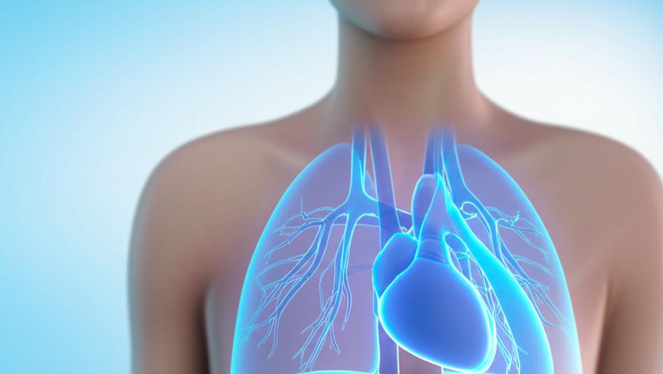
You know, it’s funny. We talk a lot about the heart as this amazing pump, and it absolutely is. But sometimes, the unsung heroes, the little pathways that make it all possible, get overlooked. I had a patient once, just curious, point to their chest and ask, “Doc, how does the good stuff, the oxygen, actually get to my heart after I breathe?” It’s a great question! And a big part of that answer lies with some really special blood vessels called the pulmonary veins. These vessels are crucial for getting that fresh, oxygen-rich blood from your lungs right to your heart, so it can be sent out to keep your whole body going.
What Exactly Are Pulmonary Veins?
So, what are these pulmonary veins? Think of them as dedicated couriers. They are blood vessels, and their main job is to carry blood that’s just picked up a fresh supply of oxygen from your lungs directly to your heart.
These veins are a key part of what we call the pulmonary circuit. This is like a special loop that moves blood between your heart and your lungs. This circuit also includes your pulmonary arteries.
Now, here’s where it gets a little different from what you might expect:
- Pulmonary arteries actually carry oxygen-poor blood from your heart to your lungs (to pick up oxygen).
- Pulmonary veins, on the other hand, carry oxygen-rich blood from your lungs back to your heart.
It’s a bit of a switcheroo from the rest of the body, isn’t it? Usually, arteries carry oxygen-rich blood away from the heart, and veins carry oxygen-poor blood towards it. Your pulmonary veins are the only veins in your body that are packed with oxygen-rich blood. And similarly, your pulmonary arteries are the only arteries carrying oxygen-poor blood. Weird, right?
But one thing stays consistent: veins (including pulmonary ones) always carry blood toward the heart, and arteries (including pulmonary ones) always carry blood away from the heart.
Most folks have four pulmonary veins, but some might have three or five. It’s just a little anatomical variation, like having slightly different fingerprints, and doesn’t usually cause any health issues.
The Crucial Job of Your Pulmonary Veins
Their function is simple but absolutely vital. Your pulmonary veins collect all that lovely, oxygenated blood from your lungs and deliver it straight to the left side of your heart. From there, your heart can pump this refreshed blood out to every organ and tissue in your body.
We often praise the heart for its pumping power, and rightly so! But without the pulmonary veins doing their part, the heart wouldn’t have that oxygen-rich blood to send out. So, they’re essential teammates in keeping everything running smoothly.
A Closer Look: Anatomy of Pulmonary Veins
Let’s get a bit more specific about where these important vessels are and what they look like.
Where are they hiding?
Your pulmonary veins are found nestled between your lungs and your heart. In each lung, many smaller blood vessels join up to form a pair of pulmonary veins. Each pair then leaves its lung through an area called the hilum (think of it as the lung’s root) and travels to connect with the left atrium – that’s the top left chamber of your heart.
What’s their structure?
Typically, you have four main pulmonary veins, two coming from each lung:
- Right superior pulmonary vein: Drains the upper and middle sections (lobes) of your right lung.
- Right inferior pulmonary vein: Drains the lower section of your right lung.
- Left superior pulmonary vein: Drains the upper section and a little tongue-like part (the lingula) of your left lung.
- Left inferior pulmonary vein: Drains the lower section of your left lung.
“Superior” just means above, and “inferior” means below. So, the names tell you which lung they serve and their general position.
Normally, each of these four veins connects directly to your left atrium, making four little openings (we call these ostia). The oxygen-rich blood flows through these openings into the left atrium, then moves to the left ventricle (the bottom left chamber), which then powerfully pumps it out to the rest of your body via a large artery called the aorta.
What about anatomical variations?
Our bodies are wonderfully diverse, and that includes pulmonary veins.
- Harmless Variations: About 38% of people have slight differences in how their pulmonary veins are set up. For example, sometimes the two left pulmonary veins join together to form a shared trunk before they enter the left atrium. This is called a common left-sided trunk and is the most common variation. Or, someone might have an extra (accessory) right pulmonary vein. These variations are usually perfectly fine because the oxygen-rich blood still gets to the left atrium where it needs to go.
- Variations That Can Cause Trouble: Sometimes, though, these variations mean the pulmonary veins don’t connect to the left atrium correctly. This can really affect how the heart works and can be quite serious. These are usually congenital heart defects, meaning they’re present from birth.
- Total Anomalous Pulmonary Venous Return (TAPVR): This is a serious condition where none of the pulmonary veins connect to the left atrium. Instead, they might connect to the right atrium or other veins leading there. This means oxygen-rich blood mixes with oxygen-poor blood, and the left side of the heart doesn’t get its proper supply. Babies with TAPVR need surgery. It’s diagnosed in about 1 in 7,809 babies in the U.S.
- Partial Anomalous Pulmonary Venous Return (PAPVR): In PAPVR, at least one pulmonary vein does connect to the left atrium, so some oxygen-rich blood gets to where it needs to go. While these babies might still need treatment, the situation often isn’t as immediately critical as TAPVR. This can sometimes go unnoticed until adulthood and might affect around 1 in 143 people.
Often, babies with TAPVR or PAPVR also have a small hole between the top two chambers of their heart (an atrial septal defect). This little hole can actually be a lifesaver, as it allows some blood to cross from the right atrium to the left atrium and then out to the body, keeping the baby going until they can get treatment.
How big are they?
A healthy pulmonary vein is usually between 9 to 13 millimeters in diameter. They can change a bit in width as they travel from your lungs to your heart. Generally, they get wider as they get closer to the heart, except for the left inferior one, which tends to start wider and get a bit narrower.
What are they made of?
Like other veins, pulmonary veins have three layers:
- The tunica adventitia (outer layer): Provides structure.
- The tunica media (middle layer): Contains smooth muscle that lets the vein widen or narrow.
- The tunica intima (inner layer): Lined with smooth cells so blood can flow easily.
A unique thing about pulmonary veins is that they have a little “sleeve” of heart muscle tissue (myocardial layer) covering a part of them near where they connect to the left atrium. Interesting, eh?
When Pulmonary Veins Face Challenges
While they’re usually quiet workers, sometimes issues can arise with the pulmonary veins.
Some conditions that can affect them include:
- Partial Anomalous Pulmonary Venous Return (PAPVR): As we talked about, where one or more pulmonary veins don’t connect to the left atrium.
- Pulmonary vein obstruction: A blockage in one of the veins, often due to a tumor in the heart or lungs.
- Pulmonary vein stenosis: A narrowing of one or more veins. In babies, this can be linked to congenital heart defects. In adults, it can sometimes be a complication after a procedure called catheter radiofrequency ablation (used to treat atrial fibrillation).
- Pulmonary vein thrombosis: A blood clot in a pulmonary vein, more common in people with lung cancer or after a lung transplant.
- Pulmonary venous hypertension: This is when there’s high blood pressure in your pulmonary veins. It’s often a result of problems with the left side of your heart, like left-sided heart failure, or issues like mitral valve stenosis.
- Total Anomalous Pulmonary Venous Return (TAPVR): The more serious congenital condition where no pulmonary veins connect to the left atrium.
The Link to Atrial Fibrillation (A-Fib)
You might hear about pulmonary veins in connection with atrial fibrillation (A-Fib). This is an irregular heart rhythm that often starts in the pulmonary veins themselves, or where they join the left atrium. The left superior pulmonary vein is a common starting point. To treat A-Fib, doctors sometimes perform a procedure called pulmonary vein isolation, which uses a catheter to target these areas.
Checking on Your Pulmonary Veins
If we need to take a look at your pulmonary veins, we have several ways to do it. These are usually imaging tests, and they don’t hurt:
- A Chest X-ray can give us a general picture.
- An Echocardiography (often called an “echo”) uses sound waves to create images of your heart and can show blood flow.
- An MRI (Magnetic Resonance Imaging) uses magnets and radio waves for detailed images.
- A CT scan (Computed Tomography) uses X-rays from different angles to create cross-sectional images.
We’ll always discuss which test, if any, is right for you.
Taking Care of Your Pulmonary Veins
If you’ve been told you have a condition affecting your pulmonary veins, the most important thing is to work closely with your healthcare team. Many issues with these veins are linked to other medical conditions, so managing those well is key.
Don’t hesitate to ask us questions about what you can do at home to manage your condition or any symptoms you might be experiencing. We’re here to help you understand and navigate it all.
Key Things to Remember About Pulmonary Veins
It’s a lot to take in, I know! Here are the main points:
- Pulmonary veins are vital for carrying oxygen-rich blood from your lungs to your heart.
- They are unique because, unlike other veins, they carry oxygenated blood.
- Most people have four pulmonary veins, but variations are common and often harmless.
- Some congenital conditions like TAPVR and PAPVR involve abnormal connections of the pulmonary veins and require medical attention.
- Pulmonary veins can be involved in conditions like atrial fibrillation.
- If there’s a concern, tests like echocardiography or CT scans can help us see what’s going on with your pulmonary veins.
You’re doin’ great just by learning more about how your amazing body works. Remember, understanding is the first step to taking good care of yourself. And you’re not alone in this.


