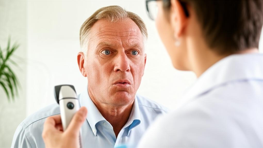
I remember a patient, let’s call him Mr. Henderson, who came into the clinic a while back. He wasn’t having any dramatic vision problems, but he mentioned his father had “that eye pressure thing,” and lately, reading the newspaper in the evening just felt… different. A bit off. It’s often that subtle feeling, or a family connection, that starts the conversation about Glaucoma Tests. And it’s a really important conversation to have, because glaucoma can be quite sneaky.
You see, glaucoma isn’t just one thing. It’s a group of eye conditions that can damage your optic nerve. Think of the optic nerve as the vital cable that sends all the visual information from your eye to your brain, allowing you to see. Most of the time, this damage is linked to an increase in pressure inside your eye. The tricky part? It often creeps up without any warning signs in the early stages. That’s why these tests are so valuable.
H2: So, What Exactly is Glaucoma?
At its heart, glaucoma is about that damage to the optic nerve. If the nerve gets damaged, it can lead to vision loss, and in serious cases, even blindness. The pressure inside your eye, what we call intraocular pressure, plays a big role. If this pressure is too high, it can slowly harm the delicate nerve fibers.
The challenging thing with glaucoma is that you might not notice anything is wrong until some vision has already been lost. It often affects your peripheral (side) vision first, and let’s be honest, we don’t always pay close attention to what’s happening at the very edges of what we see.
H2: Who Should Be Thinking About Glaucoma Tests?
Now, you might be wondering, “Is this something I need to be concerned about?” Well, some folks do have a higher chance of developing glaucoma. We often see it run in families, so if a close relative has it, your risk is a bit higher. Other things that can increase your risk include:
- Being of African or Caribbean heritage (for open-angle glaucoma, the most common type).
- Having East Asian ancestry (for closed-angle glaucoma, a less common type).
- Being very farsighted (can be a risk for closed-angle glaucoma).
- Having had a significant eye injury or complex eye surgery in the past.
- Living with diabetes.
- Having high blood pressure (hypertension).
- Long-term use of corticosteroid medications (like prednisone).
- And simply, getting older – especially after 40.
And, of course, if you start noticing any changes in your vision, that’s a definite cue to get things checked. Symptoms that might ring an alarm bell include:
- Blind spots appearing in your vision.
- Vision that seems blurry or hazy.
- Eye pain or a feeling of pressure.
- Persistent headaches, especially around the eyes.
- Seeing rainbow-colored circles when you look at lights.
- Eyes that are frequently red.
- A sense of tunnel vision, like you’re looking through a tube.
Eye specialists, the ophthalmologists, often recommend regular eye exams that include glaucoma tests, particularly if you have some of those risk factors or as you get a bit older. The American Academy of Ophthalmology, for instance, has guidelines: every one to three years for those aged 40 to 54 at risk, more frequently as you get older, or if specific concerns arise.
H2: Your Glaucoma Tests: What’s Involved?
If we think glaucoma tests are a good idea for you, you’ll typically see an ophthalmologist. They’re the eye doctors who specialize in diagnosing and treating eye conditions like glaucoma. The great news is that most of these tests are quick, generally painless, and don’t involve anything invasive. They’re all about getting a clear picture of your eye health.
So, what might they do? Here’s a rundown of some common tests:
H3: Peeking at the Angle: The Angle Exam (Gonioscopy)
This test lets the doctor look at the angle where your cornea (the clear front surface of your eye) meets your iris (the colored part). This angle is where the fluid inside your eye drains out. It’s like checking the eye’s plumbing system.
- How it’s done: Your ophthalmologist will use numbing eye drops. Then, they’ll gently place a special contact lens with mirrors on your eye. This lens allows them to see if the drainage angle is open and working well, or if it’s narrow or closed, which could be a sign of glaucoma.
H3: Measuring Up: Corneal Thickness Measurement (Pachymetry)
The thickness of your cornea can actually influence eye pressure readings and can be a factor in your overall glaucoma risk.
- How it’s done: Again, numbing drops make this comfortable. A small, pen-like probe gently touches the surface of your cornea for a moment to measure its thickness. It’s very quick!
H3: A Wider View: The Dilated Eye Exam
This is a pretty standard part of a comprehensive eye check-up. By dilating your pupils, the doctor gets a much better view of the structures at the back of your eye.
- How it’s done: Special eye drops are used to widen your pupils (the black circles in the center of your eyes). Once they’re dilated, the doctor uses a special magnifying lens and a light to look directly at your retina and, crucially, your optic nerve head, checking its shape, color, and overall health for any signs of damage.
H3: Checking the Pressure: Eye Pressure Check (Tonometry)
This is one of the most important glaucoma tests because increased eye pressure is a primary risk factor.
- How it’s done: Your eye will be numbed with drops. Then, a very precise instrument (often a tonometer) will briefly and gently touch the surface of your eye to measure the intraocular pressure. Sometimes, a puff of air is used, though direct contact tonometry is very common. It doesn’t hurt, but it helps to stay relaxed and still.
H3: Detailed Snapshots: Optic Nerve Imaging
Modern technology allows us to take incredibly detailed pictures of your optic nerve and retina. One common method is Optical Coherence Tomography (OCT).
- How it’s done: You’ll rest your chin on a machine and look into a lens for a minute or two while it scans your eye with light. Nothing touches your eye. The machine creates a cross-sectional map of your optic nerve and retinal layers, allowing the doctor to see very subtle changes that might indicate glaucoma, sometimes even before you’d notice symptoms.
H3: Mapping Your Vision: Visual Field Test (Perimetry)
This test checks for any missing areas in your field of vision, especially your peripheral (side) vision, which is often affected by glaucoma first.
- How it’s done: You’ll sit in front of a bowl-shaped machine. One eye is covered, and you’ll look at a central target. Small lights will flash at different points in your peripheral vision, and you’ll click a button whenever you see one. This helps create a map of your vision, highlighting any blind spots. It can take a little concentration, but you’re doin’ great just by focusing.
H2: After Your Glaucoma Tests: What to Expect
Once the tests are done, a few things to keep in mind:
- If your pupils were dilated, your vision will likely be blurry for a few hours, and you’ll be more sensitive to light. It’s a really good idea to have someone drive you home. Sunglasses will be your best friend!
- Otherwise, there are generally no major side effects. These glaucoma tests are very safe. Any blurriness or light sensitivity from dilation usually wears off within the day.
And the best part? In many cases, your ophthalmologist can discuss the results of most of these tests with you during the same appointment. It’s always a bit of a relief to get information quickly, isn’t it?
H2: Making Sense of Your Glaucoma Test Results
Your eye doctor will look at all the information from the different glaucoma tests together. It’s like putting pieces of a puzzle together to get the full picture of your eye health.
Here’s a general idea of what they might be looking for:
- Angle exam (gonioscopy): A narrow or blocked drainage angle can be a sign of, or risk for, certain types of glaucoma.
- Corneal thickness (pachymetry): A cornea that’s thinner than average can sometimes mean your actual eye pressure is a bit higher than the reading suggests, or it can be an independent risk factor.
- Dilated eye exam: The doctor looks for any changes in the appearance of your optic nerve – like its size, shape, or color, or any “cupping” (an indentation) which can indicate nerve damage.
- Eye pressure check (tonometry): While “normal” eye pressure can vary, readings consistently above 21 or 22 mmHg (millimeters of Mercury) are often a concern, though some people can have glaucoma with lower pressures, and others have high pressure without glaucoma (this is called ocular hypertension).
- Optic nerve imaging (OCT): These scans can detect thinning of the nerve fiber layer or other structural changes to the optic nerve.
- Visual field test (perimetry): Consistent patterns of blank or blurry spots in your vision, especially in the periphery, can indicate glaucomatous damage.
It’s not usually just one test result that leads to a diagnosis, but rather the overall pattern.
H2: If It Turns Out to Be Glaucoma: We Have Options
Okay, so what if the tests do point towards glaucoma? First, please take a deep breath. Discovering you have glaucoma can be worrying, I completely understand. But the really good news is that we have many effective ways to manage it, especially when we catch it early. Our main goal is to lower the eye pressure to prevent further damage to the optic nerve and preserve your sight.
Here are some of the common approaches:
- Medications: Often, the first step is prescription eye drops. There are several types that work in different ways to either reduce how much fluid your eye produces or help it drain better. Sometimes, pills might be used too.
- Laser treatment: Certain types of laser procedures can be very effective. For example, a laser can be used to help open up the drainage channels in your eye (trabeculoplasty) or create a tiny new hole in the iris (iridotomy) if you have angle-closure glaucoma.
- Surgery: If drops and laser aren’t enough, or in certain situations, surgical procedures might be recommended. These can involve creating a new drainage channel for the fluid (like a trabeculectomy) or implanting tiny drainage devices or shunts.
We’ll talk through all the options that are best for your specific situation, what the benefits and risks are, and what you can expect. You’re not alone in this.
H2: Take-Home Message: Key Points on Glaucoma Tests
Here are a few important things I hope you’ll remember about Glaucoma Tests:
- Glaucoma can be a “silent thief of sight,” so early detection through glaucoma tests is absolutely key.
- There isn’t just one test; a series of different evaluations helps your eye doctor build a complete picture.
- These tests are generally quick, not painful, and very safe.
- If you have risk factors like a family history of glaucoma, are over 40, or belong to a higher-risk ethnic group, it’s wise to discuss screening with your doctor or an eye specialist.
- Even if glaucoma is diagnosed, there are many good treatments available to help manage the condition and protect your precious vision.


