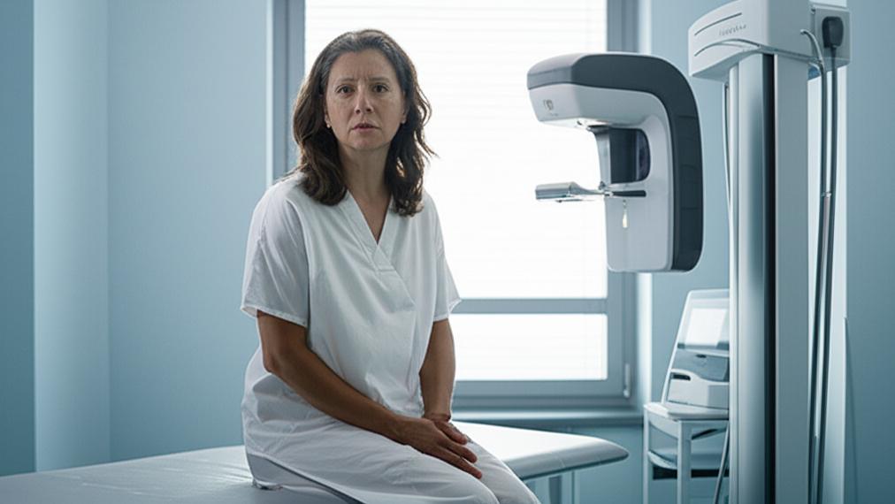
I remember a patient, Sarah, who came into my office, her face etched with worry. She’d just gotten a call back after her routine mammogram. “They said they saw something… an ‘area of concern’,” she told me, her voice trembling a little. It’s a call no one wants, and the waiting, the not knowing, can be incredibly tough. That’s often when we start talking about options like digital breast tomosynthesis, or what many know as 3D mammography. It’s a mouthful, I know – some folks say “toh-moh-SIN-thah-sis.” But what it offers can be really helpful, especially in situations like Sarah’s.
So, what exactly is this tomosynthesis we’re talking about? Think of it as a more advanced type of mammogram. A standard mammogram, which has been our trusted go-to for breast cancer screening for years, gives us 2D, or flat, pictures of the breast. Tomosynthesis, on the other hand, takes many X-ray images from different angles as it moves in an arc over the breast. Then, a computer puts these images together to create a 3D-like picture. This can give us a much clearer view, almost like looking through the pages of a book instead of just at the cover.
The U.S. Food and Drug Administration (FDA) gave it the green light back in 2011, and honestly, it’s proving to be a game-changer.
Why Might Tomosynthesis Be a Good Idea for You?
Now, you might be wondering, “Is this something I should consider?” Well, anyone who needs a mammogram could potentially benefit, but it’s particularly useful if you have dense breast tissue. About half of all women do!
Let me explain a bit about dense breasts. When we look at a mammogram, fatty tissue appears dark, while dense tissue (which includes glands and fibrous tissue) looks white. The tricky part? Cancerous tumors also show up as white. So, in dense breasts, it can sometimes be like trying to find a snowball in a snowstorm on a standard 2D mammogram.
Tomosynthesis helps us see through that “snowstorm” a bit better. Because it takes pictures from multiple angles, it can:
- Uncover cancers that might be hidden in dense tissue.
- Help us realize that something that looked suspicious on a 2D mammogram is actually just normal overlapping tissue. This is a big one because it means fewer “false alarms” – those calls that bring you back for more tests when everything is actually okay. Fewer unnecessary worries, and I’m all for that.
When Do We Use Tomosynthesis?
We use tomosynthesis in a couple of key ways:
- Screening: The US Preventive Services Task Force generally recommends women start mammogram screenings at age 40. If you have a strong family history of breast cancer, we might start even earlier. Tomosynthesis is a great screening option because it can help find cancer early, often before you’d even notice any symptoms.
- Diagnostic: If you’re experiencing symptoms like breast pain, a new lump, or skin changes on your breast, we might use tomosynthesis to get a closer look. It helps us figure out if we need to investigate further, maybe with a biopsy (where a small sample of tissue is taken for examination).
Understanding the Tomosynthesis Procedure
If you’re scheduled for tomosynthesis, it helps to know what to expect. It’s very similar to a standard mammogram experience.
Who’s Involved?
A specially trained radiologic technologist or mammographer will be right there with you. They’ll position you for the scan and operate the machine. Later, a radiologist – a doctor who specializes in interpreting medical images – will carefully examine your images and send a report to me, or your referring doctor.
How Does It Actually Work?
Your breast will be gently positioned on a special platform. Then, a clear plastic plate will come down to compress your breast. I know, this is the part no one looks forward to, but it’s really important. Compression spreads out the tissue, which gives us a clearer picture and actually allows for a lower X-ray dose.
The main difference you’ll notice with tomosynthesis is that the X-ray arm of the machine will move in a slow arc over your breast, taking those multiple images as it goes. It’s pretty quick.
Getting Ready for Your Test
A little preparation can make things go smoothly:
- Chat with your doctor: Make sure we know your full medical history, any breast conditions, surgeries, recent vaccines, or hormones you’re taking. Family history of breast cancer is important too. And, of course, let us know if you might be pregnant or are breastfeeding.
- Previous mammograms: If you’ve had mammograms at a different clinic, try to have those images sent over. Comparing old and new images is super helpful for the radiologist.
- Timing is key: If your breasts tend to get tender around your period, try to schedule your tomosynthesis for the week or two after your period ends. You’ll likely be more comfortable.
On the Day of Your Tomosynthesis
The whole thing usually takes about 30 minutes.
- Skip the products: Don’t use any deodorant, antiperspirant, powders, lotions, or creams on your underarms or breasts that day. These can show up as white spots on the images and confuse things.
- Dress comfy: Wear a two-piece outfit if you can, as you’ll need to undress from the waist up and wear a gown.
- Jewelry: It’s best to leave necklaces and dangly earrings at home, as they can also interfere with the images.
What Happens During the Scan?
- The technologist will help you get positioned.
- Your breast will be compressed. It might feel a bit tight or uncomfortable – that’s normal. But if it’s truly painful, please let the technologist know. They can often make adjustments.
- You’ll need to stay still and may be asked to hold your breath for a few seconds while the machine takes the pictures.
- The X-ray tube moves in that arc, capturing the images.
- Then, they’ll repeat the process for your other breast.
What About Radiation?
This is a question I get a lot. Yes, tomosynthesis uses X-rays, which means a small amount of radiation. The FDA has approved its use, and the total radiation dose for a tomosynthesis scan (often done alongside a 2D mammogram or with 2D images created from the 3D data) is still very low and considered safe. Newer machines can even create the 2D views from the 3D data, making the dose only slightly higher than a standard 2D mammogram. We always use the lowest possible dose to get the high-quality images needed for accurate detection.
After Your Tomosynthesis: Results and Next Steps
A radiologist will review your images, often comparing them to any previous ones you’ve had. They’re looking for any signs that might suggest breast cancer.
What Might the Results Show?
- Good news! Everything looks clear, and there are no signs of cancer.
- Sometimes, the radiologist might see an area that needs a closer look. This doesn’t automatically mean cancer. It might mean you need more detailed mammogram views, a breast ultrasound, an MRI, or possibly a biopsy. A biopsy is the only way to know for sure if cancer cells are present.
When Will You Get Your Results?
Usually, you’ll hear back within a few days, but it can sometimes take a bit longer. Before you leave your appointment, it’s a good idea to ask how you’ll receive your results and when you can expect them.
A Few More Things About Tomosynthesis
- Is tomosynthesis better than a standard mammogram? For women with dense breasts, it often provides a clearer picture, leading to better cancer detection and fewer false positives. Research is still ongoing to see if it’s definitively better for all women for general screening, but it’s a fantastic tool in our toolkit.
- Is tomosynthesis the same as a 3D mammogram? Yes, exactly! “Tomosynthesis,” “breast tomosynthesis,” “digital breast tomosynthesis (DBT),” and “3D mammogram” all refer to the same advanced imaging procedure.
- Is it really better for dense breasts? Absolutely. That’s one of its biggest strengths. It helps the radiologist distinguish between normal dense tissue and potentially concerning areas much more effectively.
Key Takeaways on Tomosynthesis
Alright, let’s quickly recap the main points about tomosynthesis:
- It’s an advanced type of mammogram, also called 3D mammography, that creates a more detailed, 3D-like image of your breast.
- It’s especially good for women with dense breast tissue, as it can improve cancer detection and reduce false alarms.
- The procedure is very similar to a standard mammogram, involving breast compression and low-dose X-rays.
- It can be used for both routine breast cancer screening and for investigating breast symptoms.
- Talk to your doctor to see if tomosynthesis is the right screening choice for you.
Remember Sarah? After her tomosynthesis, we got a much clearer picture. The “area of concern” turned out to be just overlapping healthy tissue. The relief on her face was immense. It’s not always that straightforward, but having clearer information is always a step in the right direction.
You’re not alone in navigating these health decisions. We’re here to talk through all your options and make sure you feel informed and comfortable.


